Predict the position and stability of water molecules combined with check points
Introduction
With the rapid development of structural biology, a large number of protein structures closely related to diseases have been resolved, which greatly promotes the development of structure-based drug design (SBDD). The iteration of compound structure optimization using SBDD has played an irreplaceable role in the process of drug development.
Water molecules widely exist in drug target proteins and make important contributions to the interaction between drugs and target proteins. In drug design, replacing water molecules and interacting with water molecules is an effective method to optimize the structure of compounds. In recent years, a series of cases have emerged to improve the binding activity of compounds, enhance the selectivity of compounds and improve their pharmacokinetic properties by rationally replacing water molecules.
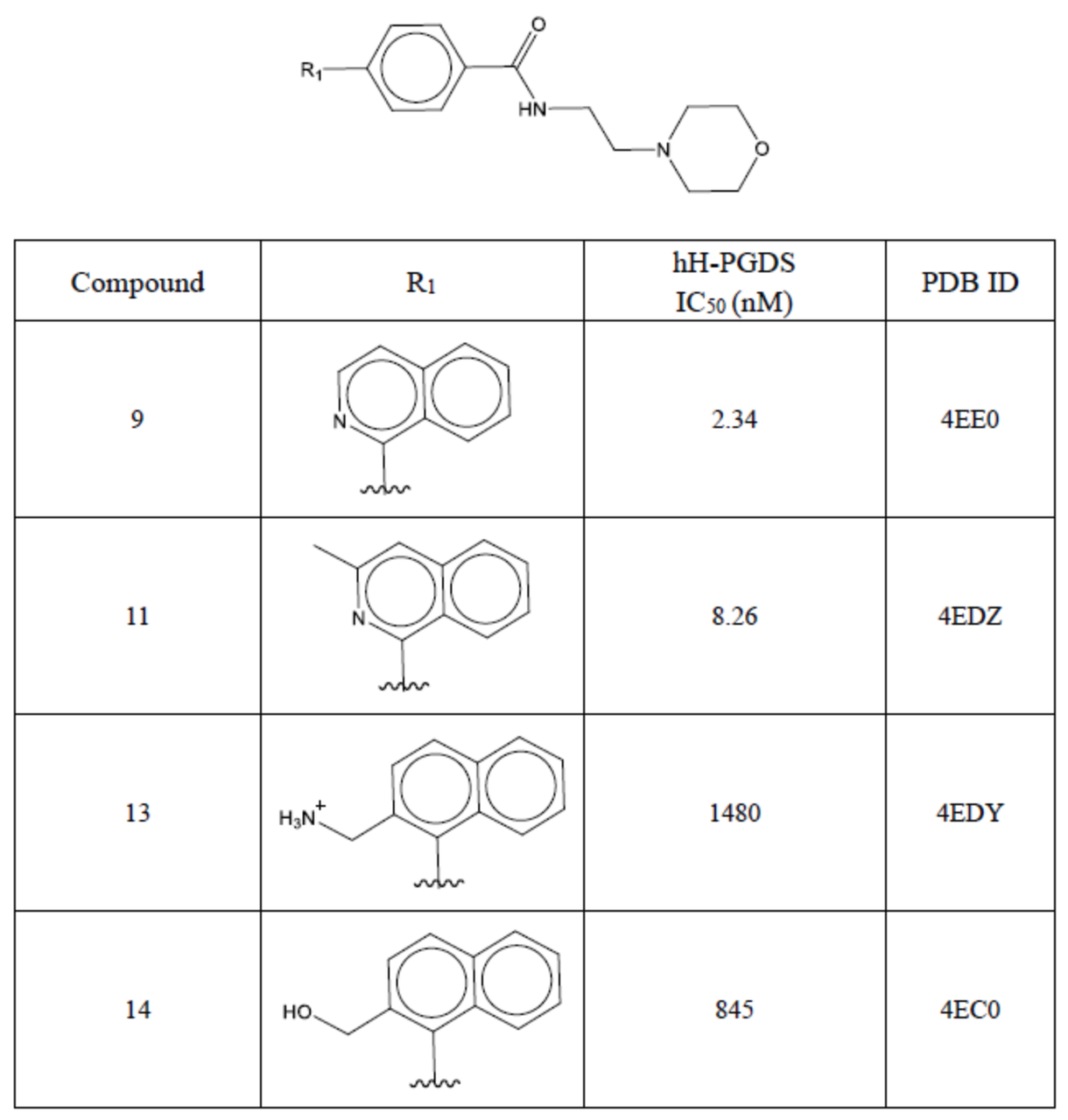
Trujillo [1] et al. explored two key water molecules (called primary water and auxiliary water in the literature, shown in this tutorial) by designing a list of hH-PGDS inhibitor lead compounds (compounds 9, 11, 13, 14). Examples of the change trend of compound affinity after substitution and substitution of water 1, water2). The experimental results of Trujillo et al. Conclusion: After replacing the auxiliary water, the activity of compound 11 decreased by 2 to 3 times compared with compound 9; after replacing the primary water, the activity of compounds 13, 14 decreased by 2 to 3 times compared with compound 9. Hundred times. This shows that primary water is more thermodynamically stable than auxiliary water.
| Four main compounds structure and activity. |
|---|
 |
| Description | 3D display |
|---|---|
| Eutectic binding mode of compound 9 with hH-PGDS (PDB 4EE0). In the binding check point, there are two water molecules Water 1 and Water 2 with hydrogen bonding networks. | 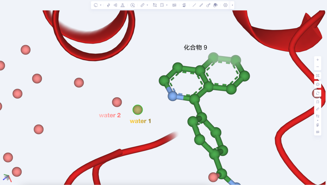 |
| Eutectic binding mode of compound 11 with hH-PGDS (PDB 4EDZ). Compound 11 introduces a methyl group to replace water 2 at the ortho position of pyridine nitrogen in compound 9, and the activity decreases by 3.5 times. | 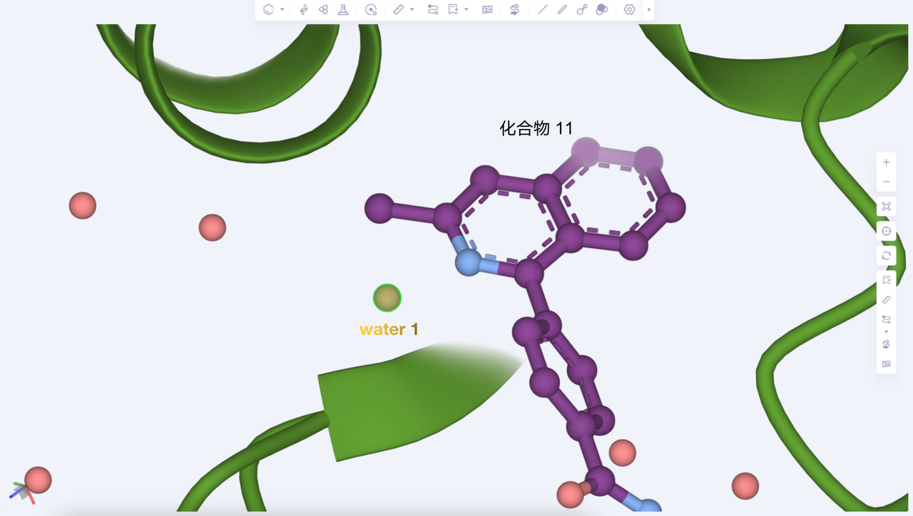 |
| Eutectic (PDB 4EDY) binding mode of compound 13 with hH-PGDS. Compound 13 replaces pyridine ring nitrogen with carbon in compound 9, and introduces amine methyl group to replace water 1, and the activity decreases 630 times. | 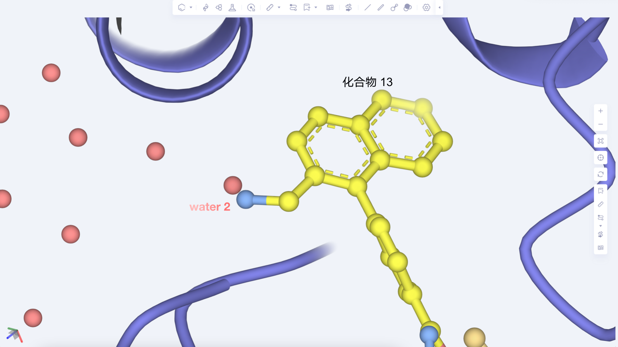 |
| Eutectic binding mode of compound 14 with hH-PGDS (PDB 4EC0). Compound 14 replaces pyridine ring nitrogen with carbon in compound 9, and introduces hydroxymethyl group to replace water 1, and the activity decreases 360-fold. | 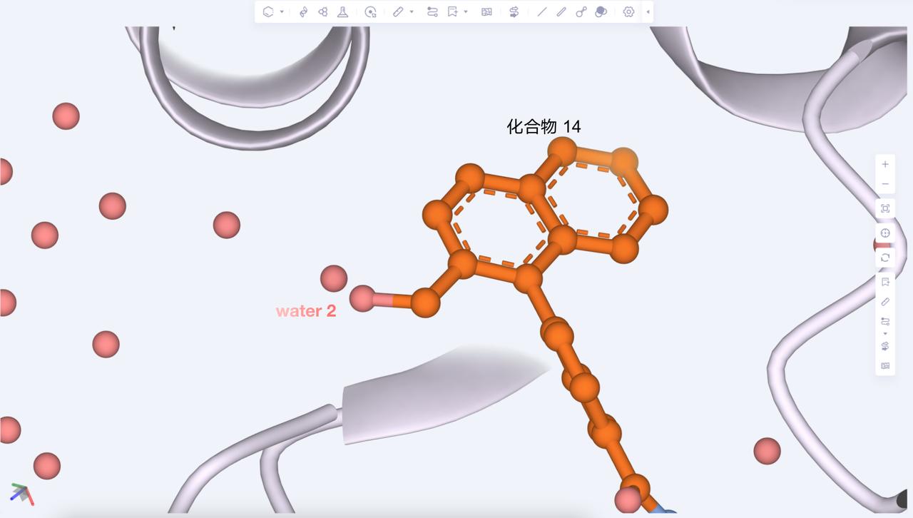 |
The purpose of this tutorial is to use Aquasite to focus on the accuracy of position prediction of two key water molecules, water 1 and water 2, and to evaluate the stability and importance of water 1 and water 2 by calculating the free energy difference of water molecules, so as to provide guidance for drug molecules. Design provides guidance.
1. Create a project and import the structure
1.1 Login system
- Login address: https://hermite.dp.tech
1.2 Create a project
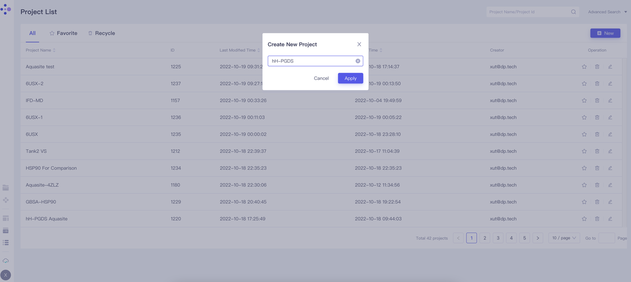
- After entering the system, create a new project "hH-PGDS"
1.3 Importing protein structures
| Operation | Display |
|---|---|
| Select File → Get PDB | 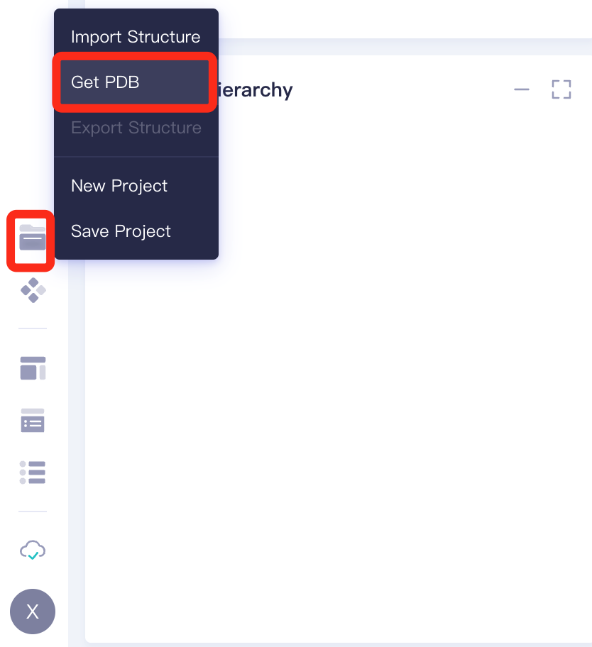 |
| Enter 4EE0 to import this protein | 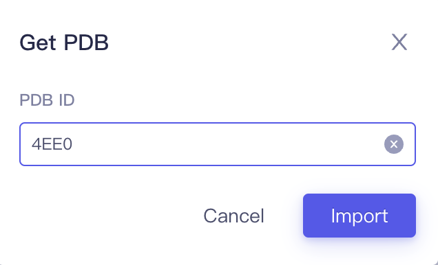 |
2. System preparation
2.1 Manual preparation system
| Operation | Display |
|---|---|
| In the Structure Hierarchy: 1. Select "Protein" Chain A, delete; 2. Select "Ligand" A 004 202, A GSF 203, B GSF 202, delete; 3. Select "Others" Metal/Ions, delete | 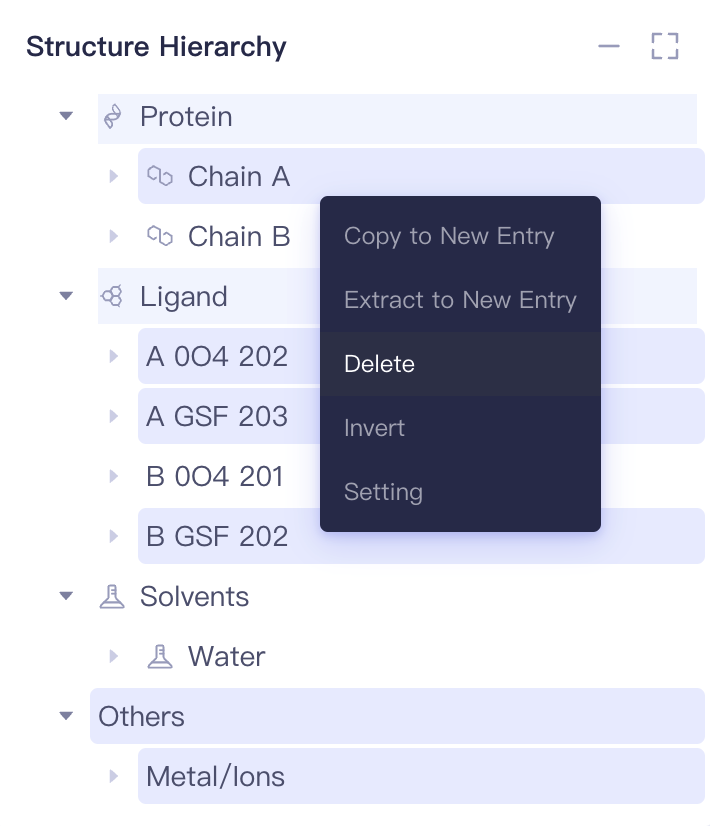 |
| In the Structure Hierarchy, keep B HOH 311, B HOH 322, B HOH 330, B HOH 337, B HOH 344, B HOH 353, B HOH 362, B HOH 372, B HOH 427, B HOH 430, B HOH 453, delete the rest of the water molecules. | 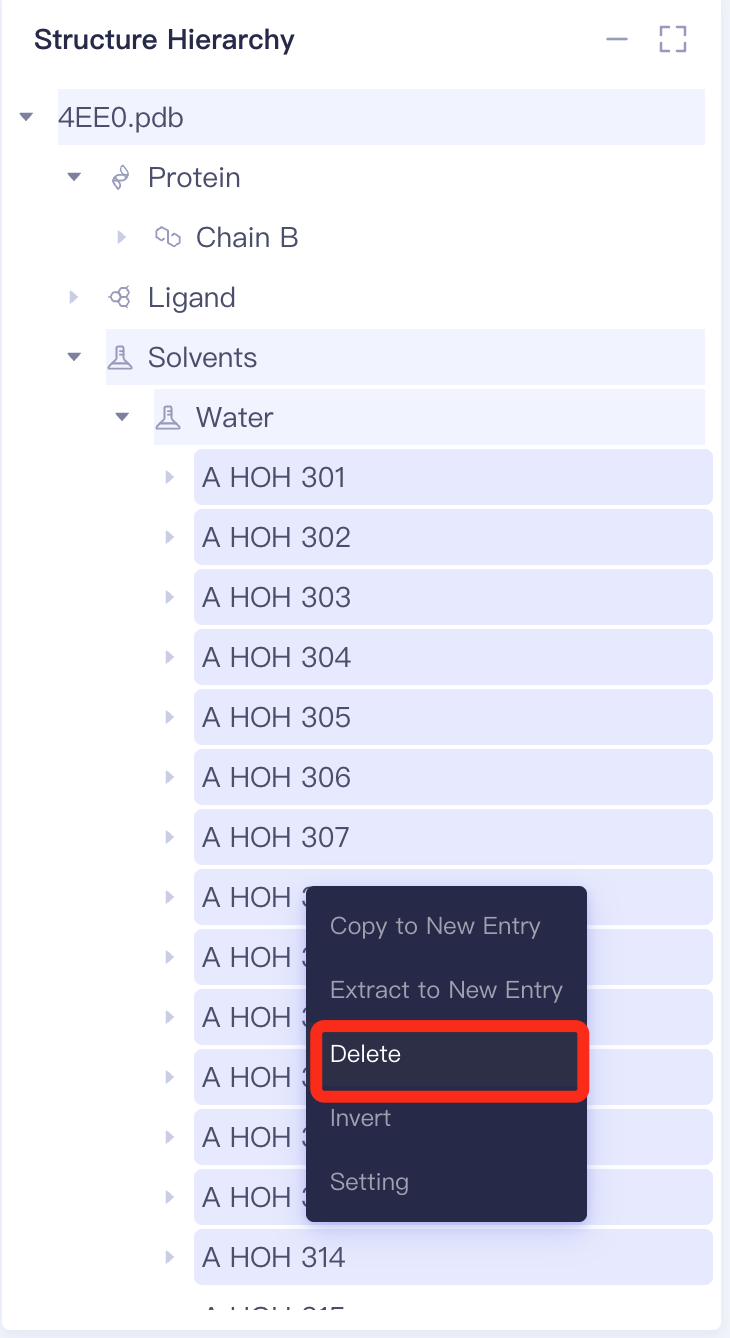 |
| After deletion, the Structure Hierarchy and protein structure are shown on the right: | 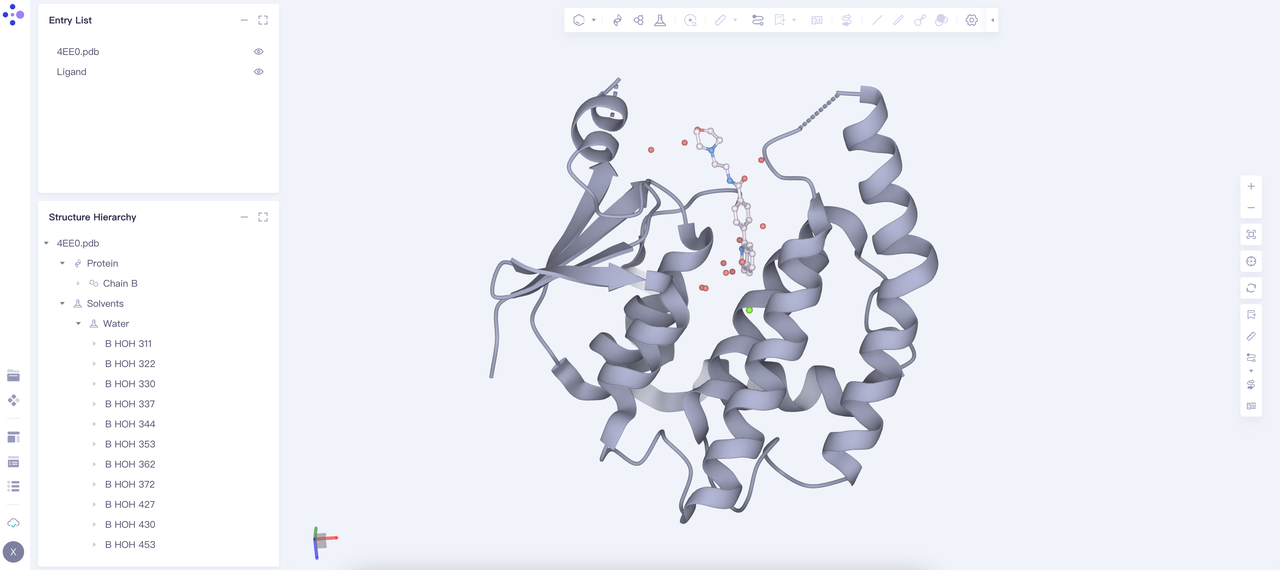 |
2.2 Extract eutectic ligand as reference molecule
| Operation | Display |
|---|---|
| Eutectic ligand B 0O4 201 "Extract to New Entry" | 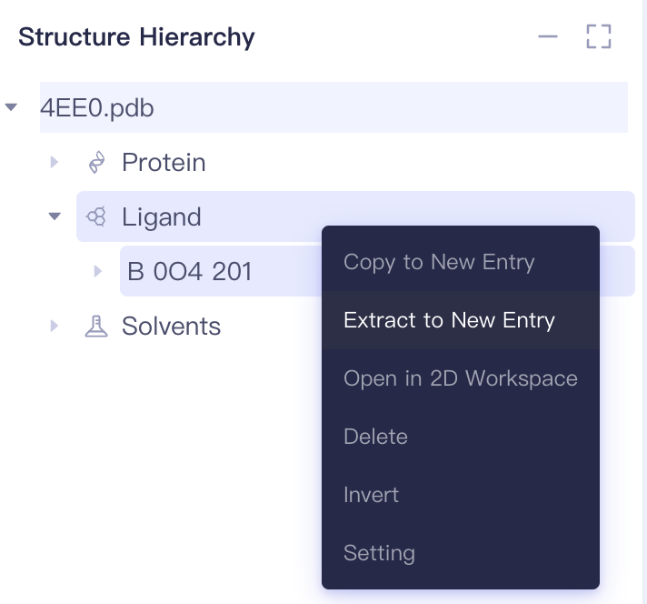 |
| Rename the Entry lists to "Ligand" | 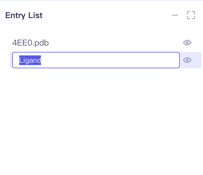 |
2.3 Prepare protein
2.3.1 Select Structure
| Operation | Display |
|---|---|
| Select Function → Structure Modeling → Protein Preparation | 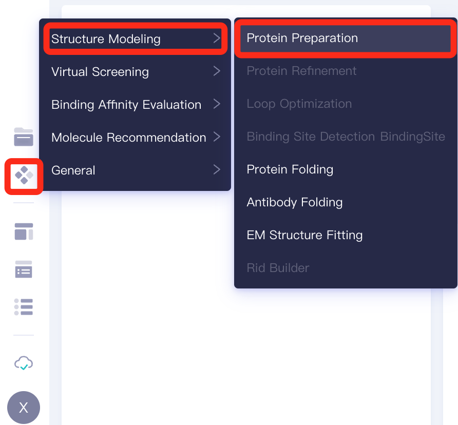 |
| Select Structure from 3D Workspace | 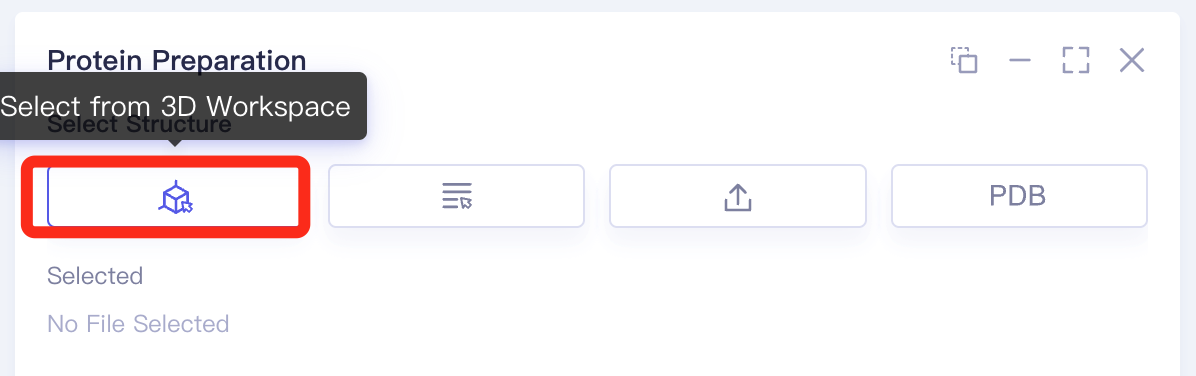 |
| Select 4EE0.pdb in the Structure Hierarchy, and in the "Select Structure" window on the right, click "OK" | 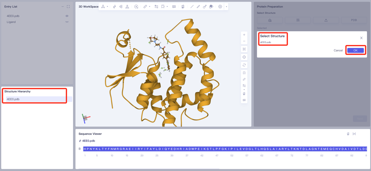 |
| 4EE0.pdb will be loaded into the parameter setting panel, click "Next" | 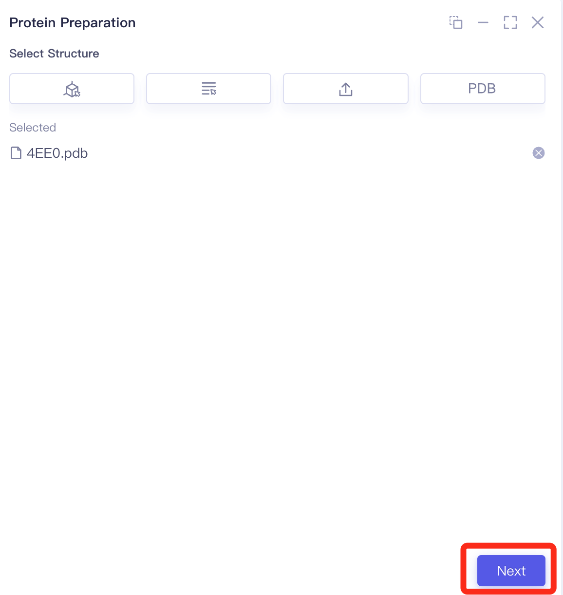 |
| Operation | Display |
|---|---|
| 2.3.2 Select Polymer, Other Groups to Keep Set according to the parameters in the figure on the right, and click “Next” | 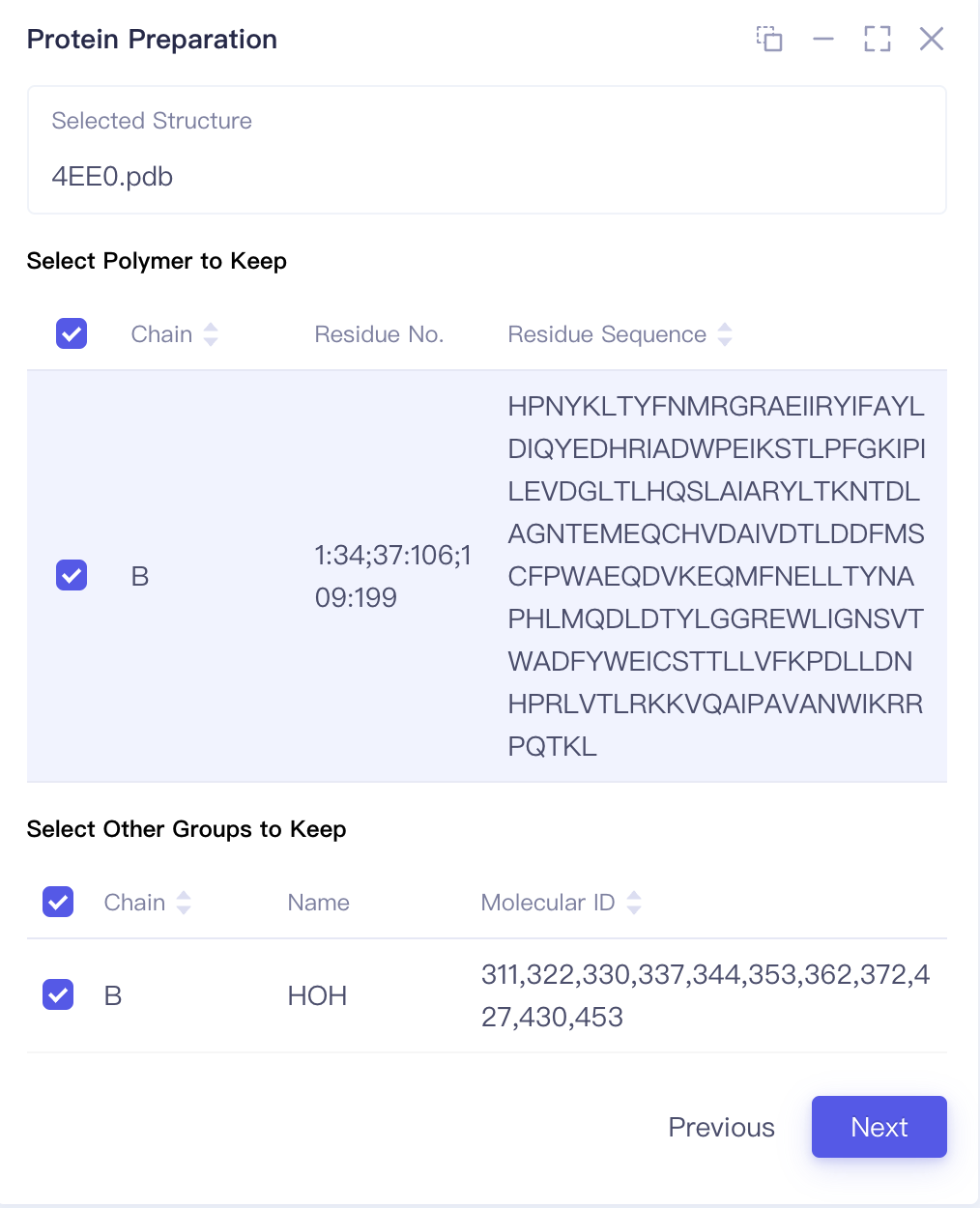 |
| 2.3.3 Select Missing Residues to Repair There is no Missing Residues for the crystal structure in this case 2.3.4 Prepared Settings Set the parameters according to the parameters in the figure below 2.3.5 Name the Job and submit the task Name the Job as "hH-PGDS-Protein Prepare", click “Submmit” to submit the task. | 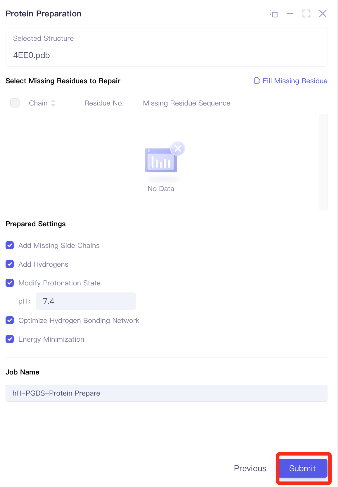 |
| After the task is submitted successfully, the 3D workspace will show the right image prompt |  |
| 2.3.6 Calculation Task View The calculation time of Protein Preparation is generally completed within ten seconds to a few minutes. After the task is over, the 3D Workspace window will automatically pop up the following message bar. |  |
| Tips Users can view the corresponding task through Jobs, and click "Show" to display the prepared protein structure in 3D Workspace. | 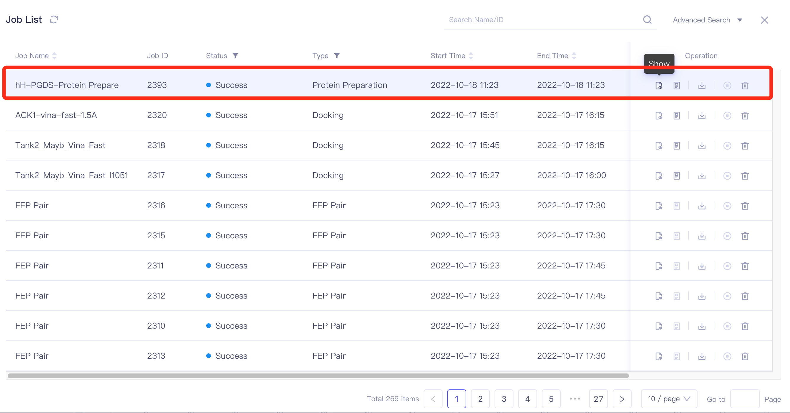 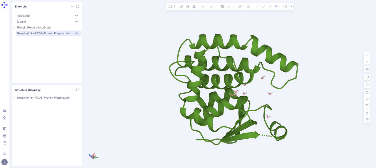 |
3. Aquasite Task Settings
3.1 Select a Prepared Protein From Project
| Operation | Display |
|---|---|
| Select General → Aquasite | 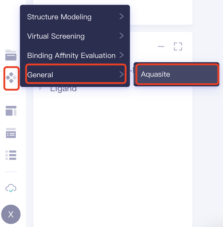 |
| Select from Project |  |
| Select "4EE0_prepared" as the prepared receptor structure, click "OK" | 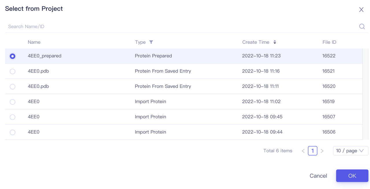 |
| After clicking "OK", the system will automatically check whether the input protein meets the calculation requirements, and the status is "Processing". | 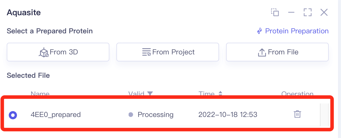 |
| In about 1 minute, the system will judge the protein as "Valid". Click "Next" | 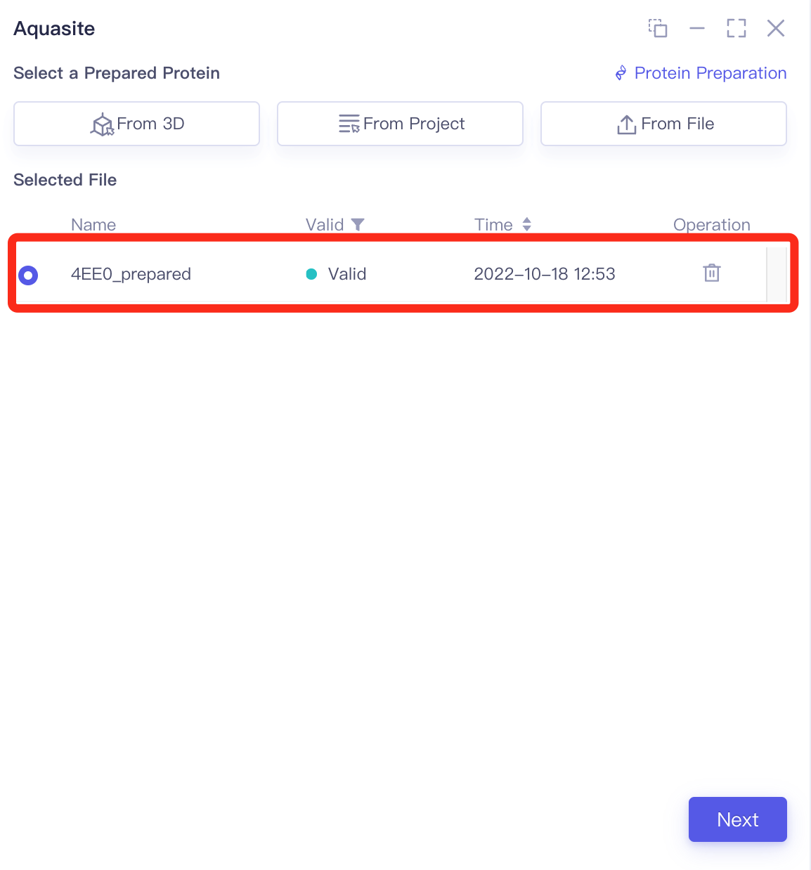 |
3.2 Select a Prepared Reference Ligand
| Operation | Display |
|---|---|
| Click "Select from 3D" | 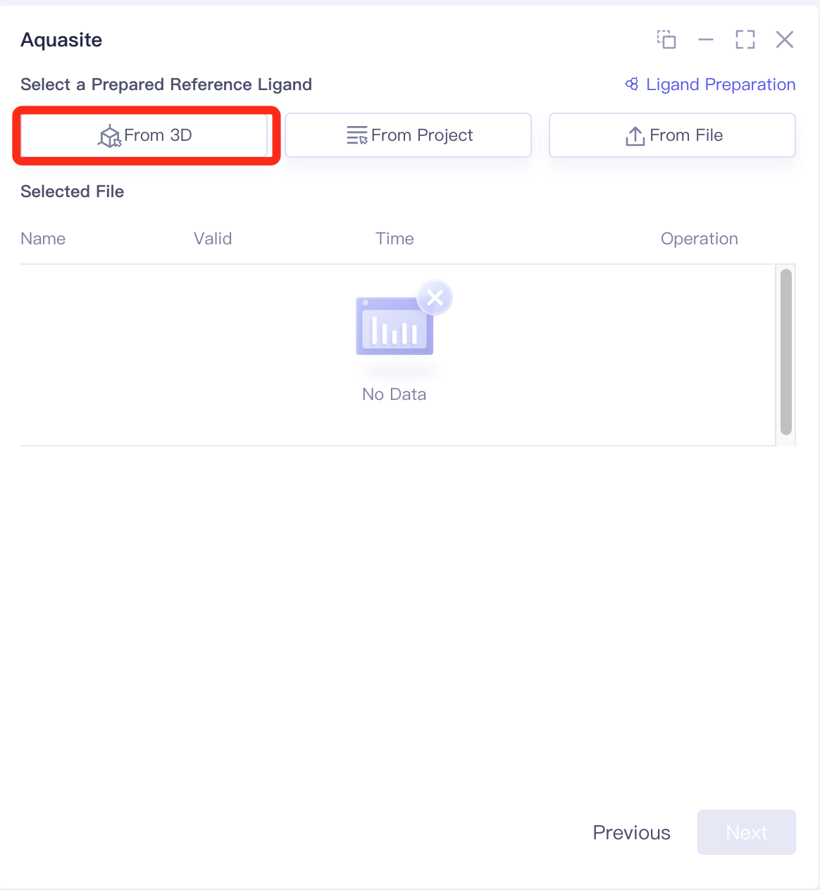 |
| Select "Ligand" in "Structure Hierarchy" | 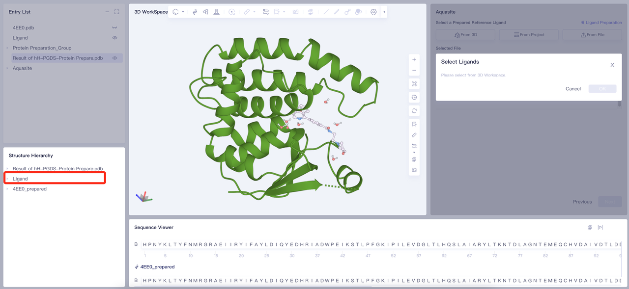 |
| Click "OK" in the "Select Ligands" window | 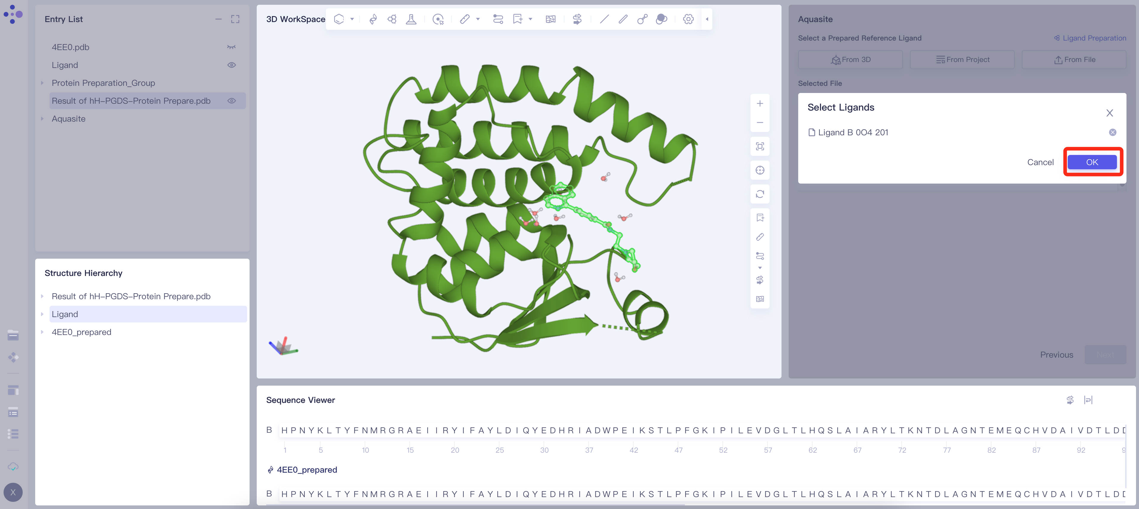 |
| Ligand_B 0O4 201 will be loaded into "Selected File". The system will automatically check whether the input Ligand meets the calculation requirements, and the status is "Processing". | 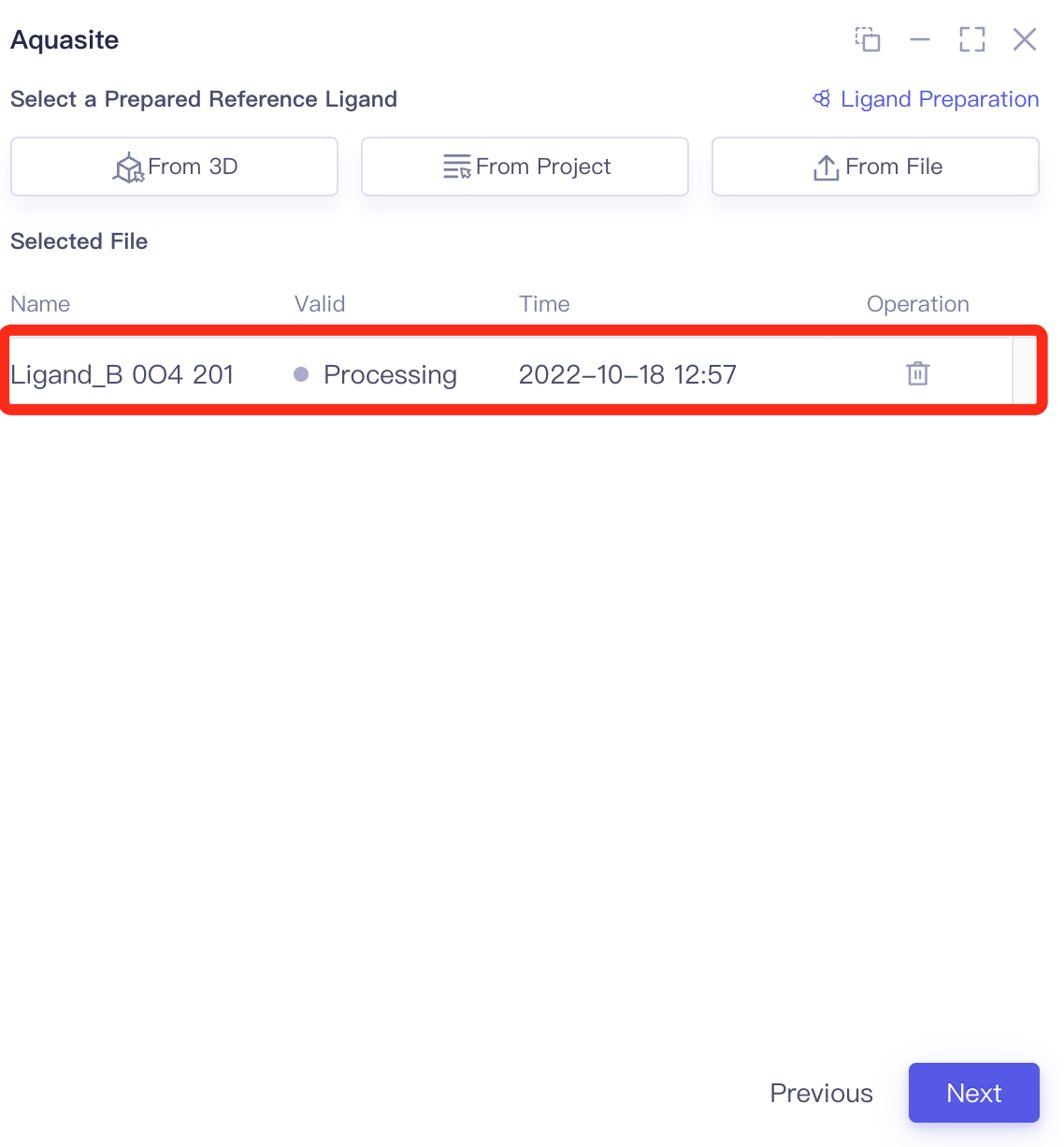 |
| In about 20 seconds, the system determines that the ligand is in the "Valid" state. | 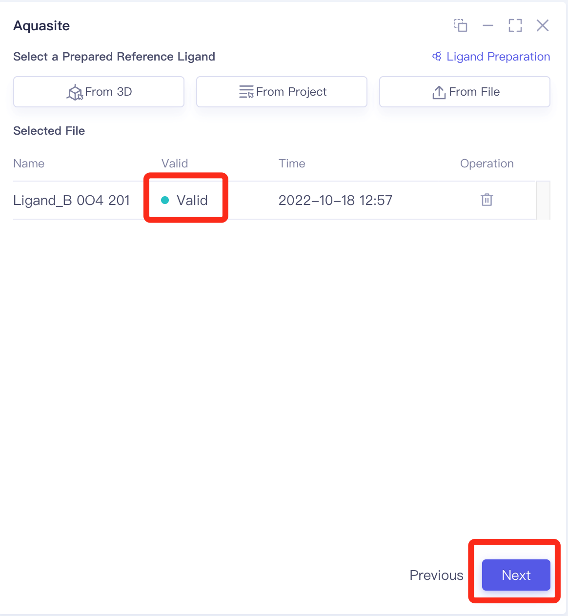 |
3.3 Confirm and Setting
| Operation | Display |
|---|---|
| Check the "Calculate with Reference Ligand" option, change the "Simulation Time" to 10 ns, and set the "Job Name" to "hH-PGDS-Aquasite". | |
| Leave the remaining parameters as default, click "Submit" to submit the calculation task. |  |
4. Analysis of Results
| Description | Display |
|---|---|
| From the Job menu on the right, open the Job List, find the "hH-PGDS-Aquasite" calculation task, and click "Show". | 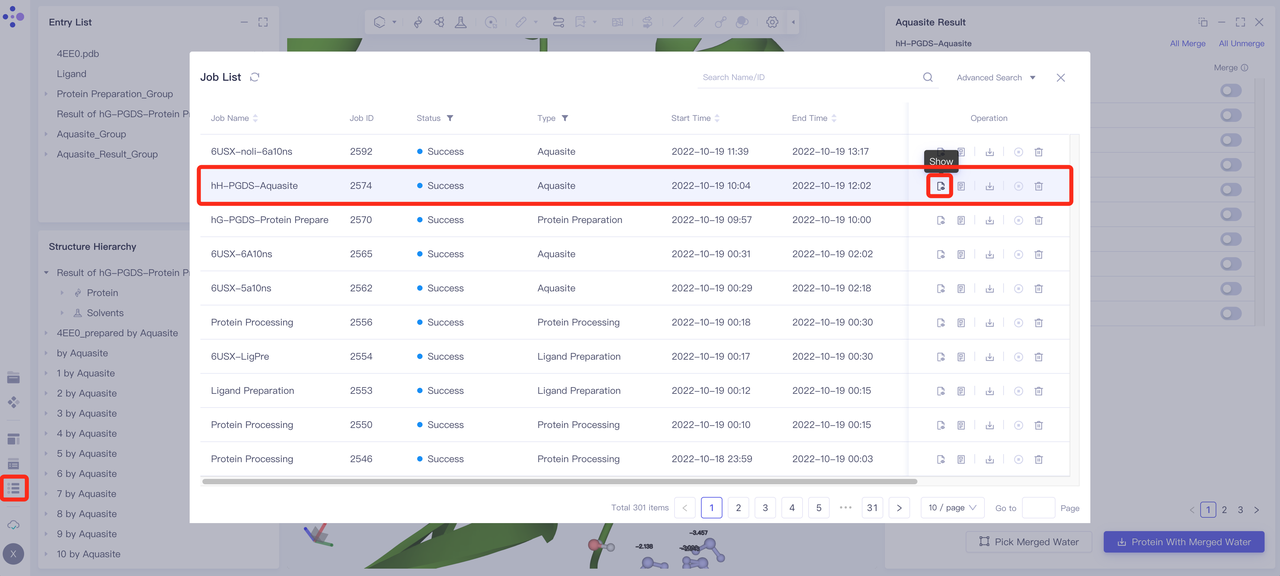 |
| 1. Adjust the molecular perspective of the 3D Workspace, focusing on showing the positions of small molecules and predicting water 1 (purple molecule in the red circle), water 2 (purple molecule in the yellow circle), water3 (purple molecule in the blue circle) and free energy. < br/> 2. From the results on the right, it can be found that the predicted positions of water 1, 2, and 3 are almost identical to the positions of eutectic water molecules in their respective labeling circles. This shows that Uni-Aquasite is relatively accurate in predicting the location precision of bound water. < br/> 3. The delta G of water 1 is -2.055 kJ/mol, and the delta G of water 2 is 0.981 kJ/mol. This shows that water 1 water molecules are more stable and more important than water 2 water molecules; substitution or substitution of such water molecules may have a significant impact on the binding affinity of compounds. This is consistent with the results reported in the literature. | 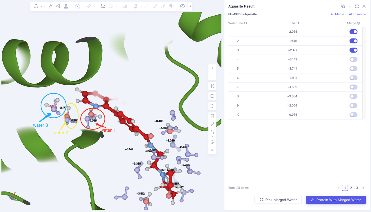 |
5. References
Trujillo, John I., et al. "Investigation of the binding pocket of human hematopoietic prostaglandin (PG) D2 synthase (hH-PGDS): A tale of two waters." Bioorganic & medicinal chemistry letters 22.11 (2012): 3795-3799.